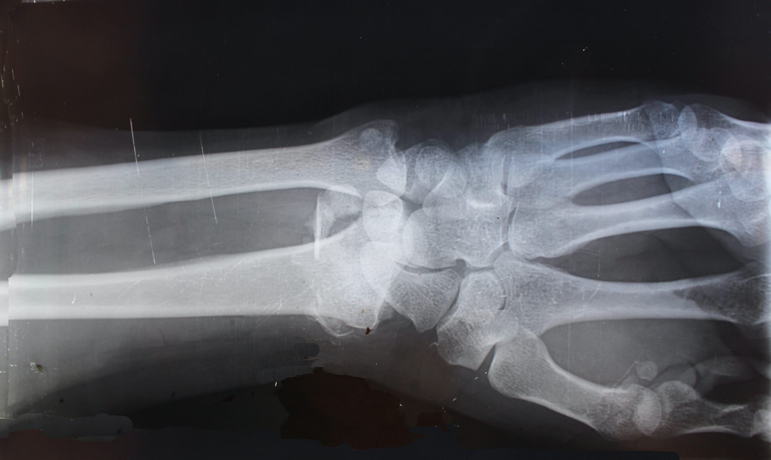
X-rays and their classification
After the stabilization, patient’s clinical status should be established. Then to identify the type of fracture, minimum two x-rays of the glenohumeral joint should be taken at right angles to each other are required. Three x-rays of the trauma-series will be better particularly for preoperative planning of B and C fractures.
If there is any susceptibility of tuberosity fractures, x-rays in external and internal rotation of the humerus may be useful. If these standardized x-rays do not help in proper assessment of the features of main fracture, displacement of fragments, of articular damage, and soft-tissue involvement then CT will be useful. To classify the fracture, and to decide the surgical procedure or prognosis, it will be necessary to determine whether the fracture runs through the anatomical or surgical neck.
Objective indications for surgery
Surgical intervention is determined by some specific factors such as: the type and stability of the fracture, general and local concomitant injuries, the age of patient and general condition and the quality of the bone (osteoporosis). Generally, the stability and displacement are interdependent. If the damage to the adjacent soft tissues and the periosteum is greater, the requirement of internal fixation and early functional post-operative treatment will also be more. Generally, in case of elderly patients, conservative treatment is given as in about 80% of cases the fracture fragments are held together by muscles, tendons, the attachment of the rotator cuff, and the periosteum. In remaining 20% reduction andoperative fixation may be required. This mainly includes cases of young patients with fractures in which more than 5 mm tuberosities are displaced and more than 2 cm shaft fragments are displaced or in which displacement of head fragment is more than 40°.
Subjective indications for surgery
One of the most important things in future decision-making is patient’s expectations. In case of elderly patients, some expects to resume sporting activities such as swimming, golf, sailing or skiing; however, others just want to be able to resume their work of everyday lives. While, restoration of preinjury levels of function must be the goal of treatment in case of younger patients.
Implants like Locking Proximal Plates and hand fracture plates are proving helpful for a surgeon to fix complex injuries in a stable manner.
Surgical Anatomy
However, differentiation of the anatomical and surgical neck fractures is difficult. Usually, the blood supply to the main head fragment in anatomical neck fractures gets disrupted and there is possibility of avascular necrosis (AVN). On the other hand, the blood supply to the head is normally preserved in surgical neck fractures.
As far as orientation between the greater and lesser tuberosities is concerned, the tendon of the long head biceps has vital role. Additionally, in displaced proximal humeral fractures or epiphysiolysis, it may be trapped between bony fragments which can make the closed reduction unattainable. In the end, a few millimeters posterior which is parallel to the tendon runs the lateral ascending branch of the anterior circumflex humeral artery which carries the most essential blood supply for the upper part of the humeral head therefore its damage can result in AVNto this particular part of the head.
Anastomosis of the anterior and posterior circumflex humeral arteries occurs on the lateral side of the surgical neck. If these vessels ruptured, the medial aspect of the capsule is the most essential remaining blood supply, mainly to the lower part of the head. When a larger medial spike of the head-fragment is present or there are no soft-tissue connections in head fragment, AVN takes place. But then, usually within 5 years, it can collapse and deformed.
An arch is formed by the acromion, along with the coracoacromial ligament and the coracoid process. The humeral head passes and rotates under this arch. This arch limits the upwards, downwards, or sideways movement of the humeral head. Infra-spinatus, sub-scapularis, supraspinatus, and teres minor muscles are attached to the tuberosities and, in conjunction with the other surrounding muscles, thus dynamic glenohumeral function is maintained by them. Reduction and fixation of the tuberosities with their muscle insertions to their original anatomical sites as closely as possible is very important to avoid impingement


 Bitcoin
  Ethereum
  Tether
  XRP
XRP  Solana
  USDC
USDC  Cardano
  TRON
TRON  Lido Staked Ether
  Avalanche
  Toncoin